Reveal Critical Structures in the Oocyte, Improve Grading, and Enable New Discoveries
Adding Oosight™ to your lab can improve success by giving you a quantitative and reproducible method to measure biological disruption in either fresh or previously frozen oocytes. You can now select oocytes for ICSI and embryos for implantation, and use the system to help improve enucleation efficiency.
Understanding the oocyte is critical to understanding embryogenesis, and studies show that a disrupted spindle apparatus or a weakened zona pellucida in the oocyte can yield lower pregnancy rates. In fact, it has been shown that pregnancy is up to 8 times more likely when the inner zona pellucida is well-ordered.
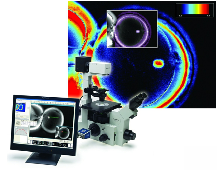
Our unique and patented solid-state, liquid crystal technology is an easy add-on to your ICSI workstation. Oosight software runs on your computer to capture, display, and analyse your images. Snap an image and click a button to report the data. Meaningful data on molecular order within the sample are organised into an intuitive, exportable report. It’s really that simple.
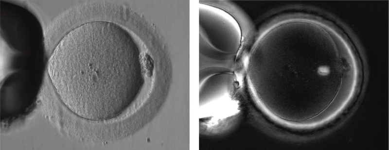
In a conventional contrast image (left) of a human MII oocyte taken just prior to ICSI, structures such as the spindle and multiple layers of the zona pellucida remain invisible. In an Oosight image (right) the spindle is clearly seen to be nicely barrel shaped and the three layers of the zona pellucida are all visible.
Oosight Imaging System is for research purposes only.